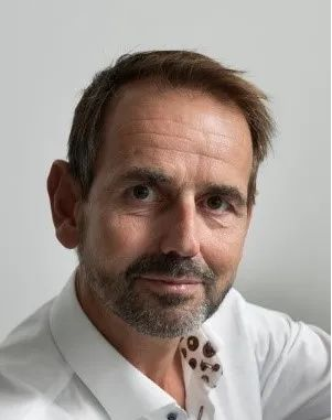
Ultrasound imaging: From ultrafast and complex to handheld-based and simple
Chris de Korte, PhD, Professor
IEEE Fellow
Medical Ultrasound Imaging Center (MUSIC), Radboudumc, Nijmegen, The Netherlands,
Physics of Fluids Group, TechMed Center, University of Twente, Enschede, the Netherlands

ABSTRACT
Ultrasound is the most versatile and low-cost medical imaging modality. It has excellent temporal and spatial resolution and allows functional imaging. With the availability of ultrafast imaging, a multitude of functional imaging techniques became available. At MUSIC, we have applied ultrafast imaging to boost 3D breast imaging, to improve strain and flow imaging in the carotid artery. Furthermore, we have developed AI-driven handheld ultrasound devices to democratize ultrasound imaging by surpassing the barrier of extensive training while minimizing the cost price.
3D breast imaging is crucial to solve the operator dependency of 2D ultrasound imaging. We developed techniques for 3D elastography using a commercially available 2D scanner. Initial evaluation reveals that malignant lesions show increased strain ratios with respect to benign types. Furthermore, we developed a cone based scanner capable of semi real-time 3D breast scanning. Additionally, ultrasensitive Doppler techniques were developed to detect neovascularization, a marker for lesion malignancy.
Atherosclerosis is the primary cause of ischemic heart failure and stroke in the westernized societies. As the natural history of atherosclerosis is associated with changes in both the vessel wall mechanics and blood flow, obtaining functional information concerning the stenosis could aid in better staging of the disease and risk stratification. Ultrafast ultrasound flow techniques where developed to quantify the complex blood flow patterns that are associated with plaque development. Additionally, dedicated coded transmission schemes where implemented to improve imaging in deeper located carotid arteries. Currently, these techniques are evaluated in 2 clinical trials. 3D strain imaging was developed and initially tested in 4 volunteers. Volumetric principle strain maps revealed locally elevated compressive and tensile strains. This new functional information could enhance our understanding of the disease process of atherosclerosis, and thereby has the potentially for earlier detection of the process of atherosclerosis.
AI driven handheld ultrasound systems where developed for non-expert users. The BabyChecker detects gestational age, position of the fetus, and single of twin pregnancy in low-resource settings with only requiring 2 hours of training for a health care worker. Evaluation in 4 African countries shows the excellent performance of this method. Additionally, a handheld- based protocol to detect hip dysplasia was developed. Using this technique, a much better detection of hip dysplasia becomes available while the healthcare workers only need 1 hour of training. These developments will revolutionize the use of medical ultrasound imaging.
BIOSKETCH
Chris L. de Korte received the M.Sc. degree in electrical engineering from the Eindhoven University of Technology, Eindhoven, Netherlands, in 1993, and the Ph.D. degree in medical sciences from the Thoraxcenter, Erasmus University Rotterdam, Rotterdam, The Netherlands, with the focus on intravascular ultrasound elastography. In 2002, he joined the Clinical Physics Laboratory, Radboud University Nijmegen Medical Center, Nijmegen, The Netherlands, as an Assistant Professor and became the Head in 2004. In 2006, he was an Associate Professor of Medical Ultrasound Techniques and he finished his training as a Medical Physicist in 2007. Since 2012, he serves as the founder and director of the Medical UltraSound Imaging Center (MUSIC), Department of Medical Imaging, Radboudumc, Nijmegen. He was appointed as a Full Professor on Medical Ultrasound Techniques in 2015. Since 2016, he has been a Full Professor on Medical Ultrasound Imaging, University of Twente, Enschede, The Netherlands. He has (co-)authored over 200 peer-reviewed articles in international journals and is (co-)inventor on four patents. His research interests include functional imaging and acoustical tissue characterization for diagnosis, treatment monitoring, and guiding interventions for oncology and vascular applications. Recently, he also focuses on AI driven apps for Point of Care Ultrasound.
Dr. de Korte is a recipient of the EUROSON Young Investigator Award of the European Federation of Societies for Ultrasound in Medicine and Biology in 1998. Furthermore he received the VENI (2000), VIDI (2004), and VICI (2010) of the Dutch Research Council NWO. In 2018, a consortium under his leadership was awarded the NWO-TTW Perspectief grant (4 million euros).
Prof. de Korte is the President of the Netherlands Society for Medical Ultrasound (NVMU) and national delegate of the European Federation of Societies for Ultrasound in Medicine and Biology (EFSUMB). He is a Fellow of IEEE. He is associate editor for IEEE Transactions UFFC and the Journal of Medical Ultrasonics. He serves as editorial board member of Ultrasound in Medicine and Biology and the Journal of the British Medical Ultrasound Society and the Technical Program Committee of the IEEE International Ultrasonics Symposium.
Time: 10:00-11:00, Sept 20, 2023 (Wednesday)
Location: Room B202, Medical Sciences Building, Tsinghua University
Tencent Meeting: 788-729-370 (Password: 202920)
Host: Jianwen Luo, PhD, Professor of Biomedical Engineering, Tsinghua University